CIRS (Now Sun Nuclear)
MRI scanners introduce geometric distortion into their images. This distortion, which can be as much as several millimeters, is problematic when the image is being used for radiation therapy or precision neurosurgical planning.
CIRS builds several grid phantoms for geometric distortion characterization. We collaborated with CIRS’s engineering team to design and develop software to characterize an MRI scanner’s geometric distortion and track it over time.
Our role on the project involved:
Through out the project we worked closely with the CIRS engineering team.
We developed an open-source fast implementation of 3D natural neighbor interpolation for this project. You can see the source code here.

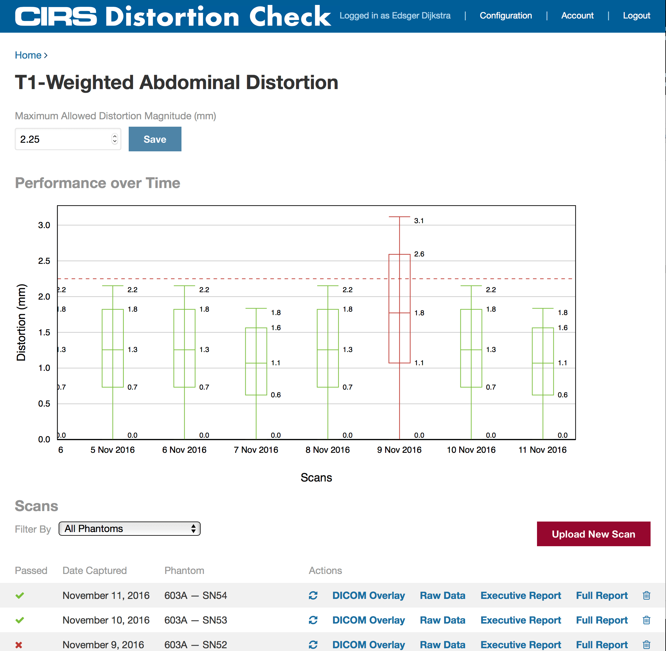

Used with MRI Grid phantoms, Distortion Check software quickly and automatically quantifies distortion in MRI images. Simply scan the phantom, upload images, review reports and trend analysis, and export DICOM overlays.
The software registers either a ground truth CAD or CT scan to the detected control points. An interpolation is then performed to generate the 3D distortion vector fields.
Results can be reported in a variety of output formats including scatter plots, contour plots, box and whisker plots for trending, and DICOM overlays that can be exported to third-party software.
Radiation Oncology
Built the dashboard, integrated with Mosaiq EMR. Still running 8 years later.
Radiation Oncology
Multi-Year Partnership: CBCT Development → FDA Clearance → Maintenance → Support → M&A Support
Every great partnership starts with a conversation. Fill out the form below for a discovery call, and an Innolitics team member will contact you soon.
I learned more from an hour-long Innolitics sales call than an entire paid engagement from other consultants.

Richard Clark
Executive in Residence at Washington University